Optical nanomanipulation for biological systems
Optical nanomanipulation for biological systems
Contact person: Dan Cojoc, G. Pinato, E. D’Este, G. Coceano, S. Finaurini, F. Tavano
|
The aim of the Optical Manipulation Laboratory (OM-Lab) is to develop new optical microscopy instrumentation and techniques for nanotechnology, biophysics and biomedicine. Optical Manipulation includes the following techniques: optical trapping and manipulation of micro and nano particles, pN force spectroscopy by optical tweezers microscopy, laser microsurgery, digital holographic and speckle microscopy, micro-Raman spectroscopy, TIRF and FRET fluorescence. Local probing the mechanical properties of living cells and local stimulation of specific compartments of neuronal cells with small amount of neuronal activation molecules represent two main directions of our research activity. Local probing of the mechanical properties employs optical tweezers (OT) force spectroscopy to measure forces exerted by membrane tether membranes on a pulling bead and derive local viscoelastic proeprties of the cell. OT are used also to locally measure cell elasticity by identation method similarly to AFM but with different loading characteristics |
Brightfield (left) and Ca ++ fluorescence images |
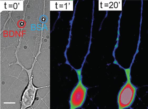
These methods are applied at OM-Lab to study the mechanical properties of cancer cells characterized by different levels of disease aggressiveness. On the other hand, digital holohraphic and speckle microscopies are employed to measure the thermal vibration of suspended cells, like red blood cells, and establish protocols to detect cell infection, e.g. malaria. Local stimulation of the neuronal cells has as goal to mimic biomechanical and biochemical interactions between hippocampal neurons, in order to understand synaptic and development mechanisms. These activities are developed in collaboration with SISSA and University of Trieste. Biomechanical interactions are mimicked using optically manipulated beads positioned in front of processes and measuring interaction forces developed there. Micro- and nano-vectors coated or filled with active molecules are instead used to stimulate specific compartments of the neurons. Examples of vectors are: microbeads, quantum dots, PLGA biodegradable beads, liposomes, micro-vesicles released by cells. The vectors are trapped and positioned on cells by optical tweezers microscopy. Stimulation is reached by cell-vector contact (coated beads or QDs), photolysis (liposomes) or by biodegradation (PLGA beasds). The effects induced are observed by optical microcopy, following the cell morphology or/and activation of specific process indicators in the cell. An example of neuronal stimulation with Brain Derived Neurothrophic Factor (BDNF) is shown in the figure above. This work (E. D’Este et al, Integrative Biology 2011) was very important since we clearly showed that BDNF not necessarily need to be internalized to trigger the receptor pathway. We also studied the effect induced by small number of molecules for two molecules of interest (Netrin and Semaphorin 3A) in neuronal signalling (G. Pinato et al, Scientific Reports 2012 ).