
Development of electron microscopy methods
Development of electron microscopy methods
Contact persons: Elvio Carlino, R. Ciancio, E. Cociancich, F. Scattarella
EMCD (electron energy loss magnetic chiral dichroism):The aim is to measure the circular dichroism by means of a TEM. Dichroism is the property of certain materials whose photon absorption spectrum depends on the polarisation of the incident radiation. Within the project it has been demonstrated the first direct experimental proof of magnetic circular dichroism in the TEM by comparing Electron Energy Loss Magnetic Chiral Dichroism (EMCD) with XMCD spectra from the same specimen together with theoretical calculations [P. Schattschneider et al. Nature 441, 486-488 (2006)]. The experiment shows that chiral atomic transitions in a specimen are accessible with inelastic electron scattering under particular scattering conditions. This result bears dramatic consequences for the study of magnetism on the nm and sub-nm scale, as EMCD offers spatial resolutions down to the sub-nanometre scale and provides depth information.
|
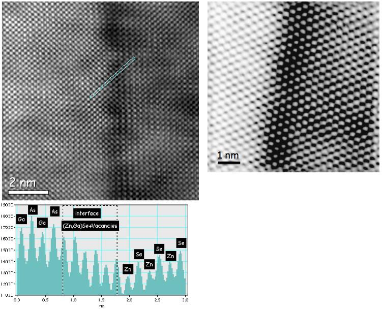
HAADF (high angle annular dark field imaging): The method (see figure) enables to image structure and chemistry of a specimen at 0.126 nm resolution. [E. Carlino et al. Phys. Rev. B 71 235303 (2005)] as contrast in the HAADF image is proportional to the atomic number of the specimen chemical species (Z-contrast imaging). The approach is based on the experiments in HAADF coupled with relevant simulation of HAADF images, by multislice calculation in the frozen-phonon framework, to derive quantitatively the correlation between experimental image contrast and the relevant chemical composition. |
HAADF image of ZnSe/GaAs [100] (left) and [110] (right) interface. The intensity profile allows to distinguish cations and anions atomic columns. |
CEDI (coherent electron diffractive imaging): relies on combining information from the high-resolution transmission electron microscopy image of an isolated nano-particle with the relevant nano-electron diffraction pattern. Phase- retrieval algorithms allow one to derive the phase, lost in the acquisition of the diffraction pattern, to visualize the actual atomic projected potential within the specimen at sub-angstroem resolution, overcoming limitations due to the electron lens aberrations. Very recently the approach has been generalized by our group to study extended crystalline specimens. The new technique has been called keyhole electron diffractive imaging (KEDI) because it aims to investigate nano-regions of extended specimens at sub-angstroem resolution by properly confining the illuminated area. In fact, by using the generalized Shannon sampling theorem it is shown that whenever suitable oversampling conditions are satisfied, EDI/KEDI diffraction patterns can contain enough information to lead to reliable phase retrieval of the unknown specimen electrostatic potential. Hence, the KEDI method has been demonstrated by simulations and experiments performed on a Si crystal cross section in the [112] zone-axis orientation, achieving a resolution of 71 pm.